Root Canal Anatomy – Mandibular Bicuspid
Managing root canal anatomy is a key element to endodontic success.
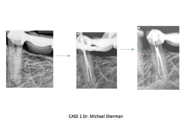
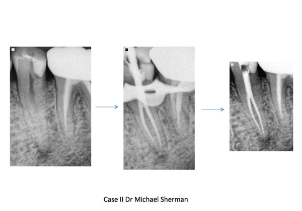
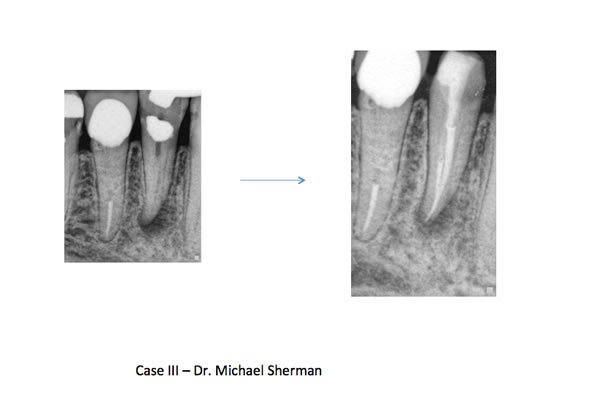
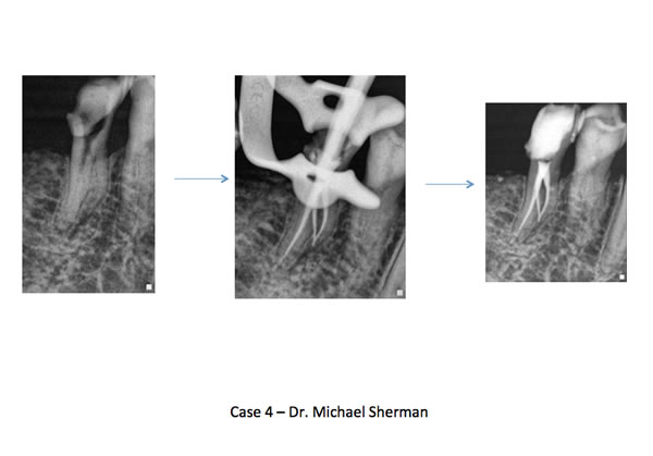
Historically, most practitioners believe the mandibular bicuspid presents with a straightforward and easy to manage root canal system. In many cases this may be true; however the mandibular biscupid may be one of the more challenging teeth we treat in our office. A mandibular first bicuspid can present with variant anatomy quite often. Zillich and Dowson reported as far back as 1973 (Journal of Oral Surgery) the presence of more than one canal nearly 23 % of the time. Two separate canals are the most common anatomical anomaly; however it is not unusual to find three canals or one canal that splits apically into multiple portals of exit. To perform successful endodontics we need to be aware of these anatomic conditions to clean and disinfect the entire canal system.
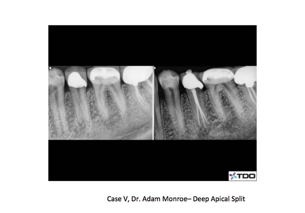
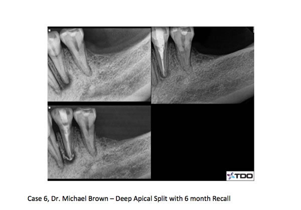
We recommend an ovoid access starting under the buccal cusp tip and extending lingual. The mandibular bicsupids can present with a lingual incline which needs to be taken into consideration when accessing under high magnification. When two canals are present, they almost always present as a buccal and lingual canal. When a deep split occurs the split usually takes place in the apical third or last several millimeters of the root. Three canals have also been reported especially in certain population subsets around the world. The mandibular first bicuspid presents with two canals more often than the second bicuspid. A well know study by Frank Vertucci in the late seventies reported the presence of two canals at the root apex nearly 25 % of the time and three canals in .5% of mandibular first bicuspids!
Preoperative awareness of additional anatomy can certainly increase detection and successful treatment of multiple canals. Examining off angle radiographs for the presence of an extra PDL can indicate multiple roots or canals. Clinically, the anatomy of the cervical area of the tooth can also hint of extra or atypical anatomy. In treating these teeth it is important to use small hand files first to explore the canal walls. A catch or deflection of the file in one direction should lead to increased suspicion of additional anatomy. Once the canals are located a thin, fine diamond can be used to flare the axial walls to ensure straight line access to each canal. I also like the use of MUNCE burs for access deep into the canal system, especially under high magnification with the micoscope. One additional complication in treating these teeth is the difficulty with obturation. Many times fitting multiple cones can be tough and can lead to “crinkling” of the cone tip. In the case of a deep split, it may be helpful to use an endodontic plugger to keep one canal patent while filling (or backfilling) an adjacent canal. I have included several cases below that were recently treated in our office. Many of these cases require more time than one would usually expect for a mandibular bicuspid. A pre-operative awareness of the manidbular bicuspid anatomy, careful instrumentation and allotting extra time will help lead to successful treatment of these cases.
I hope you enjoyed our latest endodontic blog. Please email us and share your bicuspid adventures!
Tri-City and Fallbrook Microendodontics
