Dens Invaginatus: Discovering the Possibilities…
Dens invaginatus results from an infolding of the outer surface of a tooth.
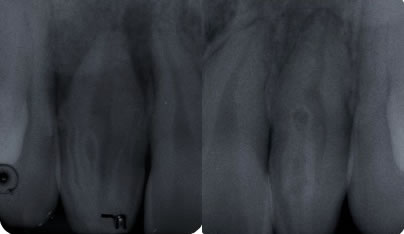
The clinical importance of dens invaginatus results from the risk of pulpal disease. All clinicians should be aware of this anomaly. The presence of quadruple dens invaginatus is extremely rare. This article presents a case of four dens invaginatus in a single permanent maxillary lateral incisor (#7) with one dens invaginatus on the opposite lateral incisor (#10). It was classified as quadruple dens invaginatus because of four enamel lined invaginations present in the crown of #7.
Dens invaginatus, known also as “dens in dente” from the literature, is a developmental anomaly resulting from invagination of enamel organ into the dental papilla, beginning at the crown and sometimes extending into the root before calcification occurs.(1,2) It commonly occurs in maxillary permanent lateral incisors followed by the maxillary central incisors, premolars, canines and less often in the molars.(1) Dens invaginatus is relatively common dental anomaly. In a review of the literature by Pindborg6 the prevalence of dens invaginatus affecting the maxillary lateral incisors ranges from 0.25% to 5.1%.
Clinically, dens invaginatus appears in the tooth crown at the site of an anatomical lingual pit susceptible to caries.(3) Radiographically, it shows a radiopaque invagination, equal in density to enamel, extending from the cingulum into the root canal.(1) The defects may vary in size and shape from a loop like, pear-shaped or slightly radiolucent structure to a severe form resembling a “tooth within a tooth”.(4) It can be identified easily because infolding of the enamel lining is more radiopaque than the surrounding tooth structure.(1)
Case Description: The 17 y.o. Asian, female patient presented to our office with a complaint of “my dentist told me I don’t have a root, that it was fused when I was born. I had pain one year ago and then again two months ago. My face was swollen and the dentist put me on antibiotics.” She was referred for teeth #6 & #7. Tooth #7 did not respond to cold, but all other teeth tested normally. Both #7 & #10 presented with dens invaginations. The lamina dura and periodontal ligament(PDL) were not intact around #7. The lamina dura was intact around #10 and the PDL was widened. A diffuse radiolucency was noted around the apices of #7, which also appeared to encroach upon #6 and #8. A widened PDL was noted around #6. Tooth #7 was diagnosed as: necrotic pulp and asymptomatic apical periodontitis. Tooth #10 was diagnosed as: normal pulp, normal periodontium.
At the initial appointment, tooth #7 was opened and several orifi were located. The canals and invaginations working lengths and sizes were identified and instrumented as the following: far mesial 15.0mmm 20/.04, main canal 18.0mm 40/.04, lingual of main canal 19.0mm 40/.04, distal 1 (blind sac) 10.0mm 30/.02, distal 2 11.0mm 30/.04. Shaping was completed with Flex-O files and GT Series X rotary files. Patency was confirmed throughout and irrigation with both 10 cc 6% NaOCl and 3 cc 17% EDTA for 30 sec in each canal and invagination using passive ultrasonic irrigation with activated with the Newtron P5.
Calcium hydroxide (UltraCal) was placed in all five canals and invaginations and temporized with a sponge and Cavit. After one month, the patient returned to re-irrigate using passive ultrasonic irrigation with the same technique listed above. GT gutta percha cones were sized using the gutta gauge and obturation was completed using the continuous wave technique with AH+ sealer, System B and HotShot. The McSpadden compactor was utilized to custom fill any voids between the two main canals. Vitrebond intraorifice barrier was placed over the gutta percha and the access sealed with Prodigy composite resin. The patient returned after two and a half years for a recall film. The lamina dura and periodontal ligament have healed.

Figure 1: Periapical radiograph showing a maxillary right lateral incisor with four dens invaginatus
Figure 2: Periapical radiograph showing a maxillary left lateral incisor with one dens invaginatus

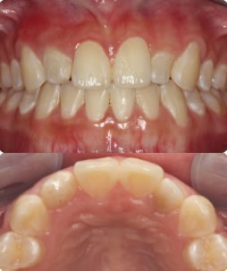
Figure 3: Facial image of maximum intercuspal position
Figure 4: Maxillary Occlusal Image

Figure 5: Image of access and orifi

Figure 6: Confirm patency

Figure 7: Periapical film – final

Figure 8: Incisal pits, Figure 9: Incisal pits prepped, Figure 10: Incisal pits bonded w/composite resin

Figure 11: Periapical film of 2.5 year recall
This tooth had an even mixture of all three types of invaginations.
Oehlers(5) described dens in dente according to invagination degree in three forms:
- Type 1: an enamel-lined minor form occurs within the crown of the tooth and not extending beyond the cemento-enamel junction;
- Type 2: an enamel-lined form which invades the root as a blind sac and may communicate with the dental pulp;
- Type 3: a severe form which extends through the root and opens in the apical region without communicating with the pulp.
She’s probably not out of the woods with tooth #10, however, the invagination does not appear to communicate with the pulp. She was placed on recall to monitor both teeth. Through a review of the literature on PubMed, I was only able to find a maximum of double invaginatus. In her case, this would be appropriately, albeit a mouthful, called a “quadruple invaginatus”.
The clinician should be aware of this anomaly because of the risk of apical inflammatory disease. Prophylactic restoration of the palatal pits of these teeth is important to avoid possible biologic injury and related inflammation.
Would taking a 3D CBCT have helped me discover this anomaly more efficiently? Most definitely! What I experienced in her case was the joy and exhilaration of discovery.
“The real voyage of discovery consists not in seeking new landscapes but in having new eyes.”
~ Marcel Proust
Thank you –
Anne Wiseman, D.D.S., M.S.D.
References
1. White SC, Pharoah MJ. Oral radiology principles and interpretation. 4. St Louis; Mosby: 2000. pp. 314–315.
2. Lee AM, Bedi R, O’Donnell D. Bilateral double dens invaginatus of maxillary incisors in a young Chinese girl. Aust Dent J. 1988;33:310–312.
3. Vajrabhaya L. Nonsurgical endodontic treatment of a tooth with double dens in dente. J Endod. 1989;15:323–325.
4. Gotoh T, Kawahara K, Imai K, et al. Clinical and radiographic study of dens invaginatus. Oral Surg Oral Med Oral Pathol. 1979;48:88–91.
5. Oehlers FAC. Dens invaginatus (dilated composite odontoma). 1. Variations of the invagination process and associated anterior crown forces. Oral Surg Oral Med Oral Pathol. 1957;10:1204–1218.
6. Pindborg JJ. Pathology of the Dental Hard Tissues. Philadelphia; WB Saunders: 1970. p. 58.
Dens Invaginatus Discovering the Possibilities…
Case by Anne E. Wiseman, D.D.S., M.S.D.
Thanks for visiting Tri City Micro Endodontics.
