Regenerative Endodontics
For this week’s blog, I want to focus on one of the hottest topics in clinical endodontics, pulpal regeneration.
In many of the latest endodontic journals there is at least one article focusing on regeneration. So what exactly is regenerative endodontics? A good definition would be a biologically based procedure designed to replace damaged tooth structure in an immature, necrotic tooth and restore pulpal function and root development (definition from Murray et al, JOE, 2008;34:S51). The loss of an immature, permanent tooth in a young patient can be very traumatic. Regenerative dentistry borrows from the principles of regenerative medicine and tissue engineering to re-establish a blood supply to a necrotic tooth. The science behind tissue engineering can be applied to endodontics and allow the retention of teeth that otherwise may be lost. In our office we have started to incorporate regenerative techniques over the past two years with success. After a discussion about the general concept of regeneration, I will run through the step by step details of the latest case performed in our office.
Regenerative endodontics stems from early research on the role of the blood clot in root canal therapy and re-establishing vascularity to existing pulp tissue. Once vascularity is re-established the root can continue development. Historically when presented with an immature, necrotic permanent tooth, we used Calcium Hydroxide over many visits in an apexification procedure to induce root closure. Multiple appointments were needed for this technique and there were negative reports on the effect of long term CAOH within the root. Over the last ten years, MTA was used to seal the open apex and create a barrier on which to condense our root canal obturation material. MTA is a wonderful material however the tooth remains truncated with thin dentinal walls. Regenerative endodontics aims to actually include root closure and re-establish a blood supply within the tooth, a feat that was not possible with either CAOH or MTA apexification. The tooth often can retain nociception and function like a normal tooth.
Today’s regeneration procedure involves a two step process. The first step aims to access and disinfect the pulp space. The second appointment focuses on stimulating growth factors from the dentin and delivering stem cells into the root through the establishment of a blood clot. I want to reinforce that this procedure is intended for young patients with underdeveloped root apices and not for closed roots in adults. I have included the protocol recommended in the article Translational Science in Disinfection for Regenerative Endodontics by Diogenes et al. published in the JOE 2014;40:S52-S57. The protocol outlined below is the technique utilized in our office with one difference. We opt to use CAOH for disinfection as opposed to the antibiotic paste. There are several articles in the current literature that report staining of the clinical crown with the A/B paste. The CAOH can be just as effective without the undesirable effect of discoloration.
The protocol is outlined below:
1. Informed consent, including explanation of risks and alternative treatments or no treatment, is obtained.
2. After ascertaining adequate local anesthesia, rubber dam isolation is obtained.
3. The root canal systems are accessed, and the working length is determined (radiograph of a file loosely positioned at 1 mm from the root end).
4. The root canal systems are slowly irrigated first with 1.5% NaOCl (20 mL/canal for 5 minutes) and then irrigated with 17% EDTA (20 mL/canal for 5 minutes), with the irrigating needle positioned about 1 mm from the root end.
5. Canals are dried with paper points.
6. Calcium hydroxide or antibiotic paste at a concentration no greater than 1 mg/mL is delivered to the canal system.
7. Access is temporarily restored.
During the final (second) treatment visit (2–4 weeks after the first visit), the following are performed:
1. A clinical examination is first performed to ensure that that there is no moderate to severe sensitivity to palpation and percussion. If such sensitivity is observed or a sinus tract or swelling is noted, then the treatment provided at the first visit is repeated. At this point, the clinician may elect to use TAP (at no more than 100 mg of each drug/mL).
2. After ascertaining adequate local anesthesia with 3% mepivacaine (no epinephrine), rubber dam isolation is obtained.
3. The root canal systems are accessed; the intracanal medicament is removed by irrigating with 17% EDTA (30 mL/canal for 10 minutes).
4. The canals are dried with paper points.
5. Bleeding is induced by rotating a precurved K-file size #25 at 2 mm past the apical foramen with the goal of having the whole canal filled with blood to the level of the cementoenamel junction.
6. Once a blood clot is formed, a premeasured piece of Collaplug (Zimmer Dental Inc, Warsaw, IN) is carefully placed on top of the blood clot to serve as an internal matrix for the placement of approximately 3 mm of white MTA (Dentsply, Tulsa, OK).
7. A (3–4 mm) layer of glass ionomer layer (eg, Fuji IX; GC America, Alsip, IL, or other) is flowed gently over the MTA and light cured for40 seconds.
8. A bonded reinforced composite resin restoration (eg, Z-100; 3M, St Paul, MN, or other) is placed over the glass ionomer.
9. The case needs to be followed up at 3 months, 6 months, and yearly after that for a total of 4 years.
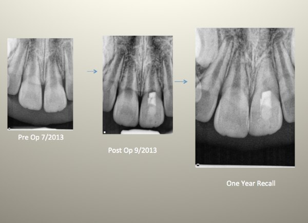
In the case below, an eight year old patient presented with pain and slight swelling localized to tooth #9. The patient had injured the tooth one year prior and the pulp eventually turned necrotic. Considering the thin dentinal walls and open apex, we opted to perform a pulpal revascularization. Following the protocol outlined above we completed the procedure and the patient’s pain resolved. The one year recall reveals further development of the root and root apex. This tooth should be less susceptible to fracture than a tooth where the apex was plugged with MTA.
Pulpal revascularization is in its infancy and I am certain there will be new developments and changes in the treatment protocol over the next decade. We hope to stay on top of the literature and will continue to post any updates as they become available.

** In writing this blog, I utilized information from the AAEs Colleague for Excellence on pulpal regeneration.
Full access to this article can be found here: http://www.aae.org/uploadedfiles/publications_and_research/endodontics_colleagues_for_excellence_newsletter/ecfespring2013.pdf
Thanks for visiting us at Tri-City and Fallbrook Micro Endoodntics of San Diego, CA.
